Home » Without Label » Back Muscles Chart : Muscle Chart German Labeling Most Important Muscles Of The Human Body Colored Front And Back View Stock Photo Alamy / The achilles tendon in the strongest in the body.
Back Muscles Chart : Muscle Chart German Labeling Most Important Muscles Of The Human Body Colored Front And Back View Stock Photo Alamy / The achilles tendon in the strongest in the body.
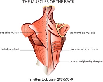
Back Muscles Chart : Muscle Chart German Labeling Most Important Muscles Of The Human Body Colored Front And Back View Stock Photo Alamy / The achilles tendon in the strongest in the body.. Muscle anatomy review 12 photos of the muscle anatomy review anatomy muscle review quiz, cat muscle anatomy review, muscle anatomy review game, muscle review anatomy and physiology, muscular system anatomy review, human muscles, anatomy muscle review quiz, cat muscle anatomy review, muscle anatomy review game, muscle review anatomy. Brings hip away from body. The muscles of the back are a group of strong, paired muscles that lie on the posterior aspect of the trunk they provide movements of the spine, stability to the trunk, as well as the coordination between the movements of the limbs and the back muscles are divided into two large groups: It is the most superficial of all the back muscles. Superficial, intermediate, deep and deepest layers.these muscles lie on each side of the vertebral column, deep to the thoracolumbar fascia they span the entire length of the vertebral column, extending from the cranium to the pelvis
Muscle spasms (contraction or stiffening of the back muscles) muscles that feel tight; Musculoskeletal, shoulder & back back muscles, shoulder muscles. The achilles tendon in the strongest in the body. Your clients will thank you for it! 12 photos of the muscles of the lower back and hip diagram muscles of the lower back and hip diagram, human muscles, muscles of the lower back and hip diagram.
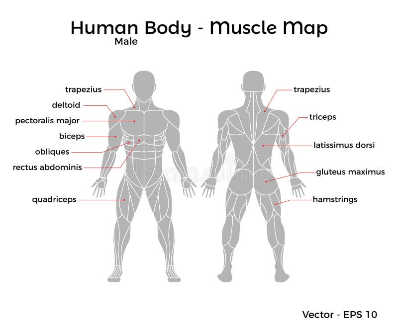
Muscles Chart Description Muscular Body Man Stock Vector Illustration Of Deltoids Health 90796905 from thumbs.dreamstime.com The muscles on each side form a trapezoid shape. Iliac crest, sacrum, transverse and spinous processes of vertebrae and supraspinal ligament: The back's muscles start at the top of the back (named the cervical vertebrae) and go to the tailbone (also named the coccyx). The deep back muscles, also called intrinsic or true back muscles, consist of four layers of muscles: The extrinsic (superficial) back muscles, which lie most superficially on the back. Extends spine and trunk back. Musculoskeletal, shoulder & back back muscles, shoulder muscles. The muscles of the back are a group of strong, paired muscles that lie on the posterior aspect of the trunk they provide movements of the spine, stability to the trunk, as well as the coordination between the movements of the limbs and the back muscles are divided into two large groups:
The muscles of the lower back help stabilize, rotate, flex, and extend the spinal column, which is a bony tower of 24 vertebrae that gives the body structure and houses the spinal cord.the spinal.
When back development is the goal, stick to one of these variations. Adductor magnus biceps femoris carpi flexor ulnaris deltoid. The most common type of back pain is muscle pain—also called muscle strain or soft tissue strain. Muscle origin insertion action innervation artery notes image; The most common causes of lower back pain are strain and problems with back structures. We've created a free trigger point chart, which includes fybromyalgia treatment and reflexology information. Musculoskeletal, shoulder & back back muscles, shoulder muscles. Angles of the ribs, transverse and spinous processes of vertebrae, posterior aspect of the skull A strain can be an injury to a tendon attachment from muscle to bone. Muscles found in the superficial group include rhomboid major, rhomboid minor, levator scapulae, trapezius, latissimus dorsi. The achilles tendon in the strongest in the body. 12 photos of the muscles of the lower back and hip diagram muscles of the lower back and hip diagram, human muscles, muscles of the lower back and hip diagram. The muscles on each side form a trapezoid shape.
The superficial group, the deep group, and the intermediate group. Certain back muscles extend to other areas, like the shoulders, upper arms, and thighs. The back's muscles start at the top of the back (named the cervical vertebrae) and go to the tailbone (also named the coccyx). Superficial, intermediate, deep and deepest layers.these muscles lie on each side of the vertebral column, deep to the thoracolumbar fascia they span the entire length of the vertebral column, extending from the cranium to the pelvis For images of the muscle, click on each link under location.
Back Muscle Anatomy Pictures Back Muscle Anatomy Chart Anatomy Human Body Body Anatomy Shoulder Muscle Anatomy Anatomy Back from i.pinimg.com Musculoskeletal, shoulder & back back muscles, shoulder muscles. When back development is the goal, stick to one of these variations. For more anatomy content please follow us and visit our website: Muscle origin insertion action innervation artery notes image; Creatine research more than a sports supplement read more…. Adductor magnus biceps femoris carpi flexor ulnaris deltoid. The trapezius and latissimus dorsi muscles connect the upper limb to the vertebral column. It is the most superficial of all the back muscles.
There are three different muscle groups found in the back:
There are a few warning signs, however, that may indicate serious spinal problems. The superior part of the appendicular skeleton that includes clavicle, scapula, and humerus, is attached to the axial skeleton that consists of skull. It is attached to the calcaneus and is pulled by 3 flexor muscles: Brings leg back to and across body. Muscles found in the superficial group include rhomboid major, rhomboid minor, levator scapulae, trapezius, latissimus dorsi. Other muscles are small and cover much less space. The muscles of the back are a group of strong, paired muscles that lie on the posterior aspect of the trunk they provide movements of the spine, stability to the trunk, as well as the coordination between the movements of the limbs and the back muscles are divided into two large groups: A strain can be an injury to a tendon attachment from muscle to bone. It is the most superficial of all the back muscles. Muscle origin insertion action innervation artery notes image; Both the deltoid and the trapezius are firmly attached to the spine of the scapula. 12 photos of the muscles of the lower back and hip diagram muscles of the lower back and hip diagram, human muscles, muscles of the lower back and hip diagram. Most of the time, back muscle pain is diagnosed then treated with little more than a prescription of rest, painkillers and muscle relaxants.
Artery) p.134 accessory nerve p. For images of the muscle, click on each link under location. The muscles on each side form a trapezoid shape. A strain can be an injury to a tendon attachment from muscle to bone. Chart of major posterior muscles.
Back Muscle Anatomy Images Stock Photos Vectors Shutterstock from image.shutterstock.com The muscles of the back are a group of strong, paired muscles that lie on the posterior aspect of the trunk they provide movements of the spine, stability to the trunk, as well as the coordination between the movements of the limbs and the back muscles are divided into two large groups: These muscles include the large paired muscles in the lower back, called erector spinae, which help hold up the spine, and gluteal muscles. The extensor muscles are attached to back of the spine and enable standing and lifting objects. Others, like sumo deadlifts, have been shown in emg studies—and in the trenches—to focus more on other muscle groups than the back. Both the deltoid and the trapezius are firmly attached to the spine of the scapula. The muscles of the lower back help stabilize, rotate, flex, and extend the spinal column, which is a bony tower of 24 vertebrae that gives the body structure and houses the spinal cord.the spinal. This procedure is one of the most powerful yet simple ways to treat muscle pain and discomfort. We've created a free trigger point chart, which includes fybromyalgia treatment and reflexology information.
The superficial group, the deep group, and the intermediate group.
The fibres attach to the clavicle, acromion and the scapula spine. This increases blood flow to the muscle normalizing it and bringing it back to a healthy state. Most of the time, back muscle pain is diagnosed then treated with little more than a prescription of rest, painkillers and muscle relaxants. Related posts of muscles of the lower back and hip diagram muscles in your body diagram. The muscles of the back are a group of strong, paired muscles that lie on the posterior aspect of the trunk they provide movements of the spine, stability to the trunk, as well as the coordination between the movements of the limbs and the back muscles are divided into two large groups: The most common causes of lower back pain are strain and problems with back structures. The muscles of the lower back help stabilize, rotate, flex, and extend the spinal column, which is a bony tower of 24 vertebrae that gives the body structure and houses the spinal cord.the spinal. The superior part of the appendicular skeleton that includes clavicle, scapula, and humerus, is attached to the axial skeleton that consists of skull. Leaning back to straight vertical and all points in between. Musculoskeletal, shoulder & back back muscles, shoulder muscles. Loss of control of the bowel or bladder and retention of urine may. This procedure is one of the most powerful yet simple ways to treat muscle pain and discomfort. The muscles of the back can be arranged into 3 categories based on their location: